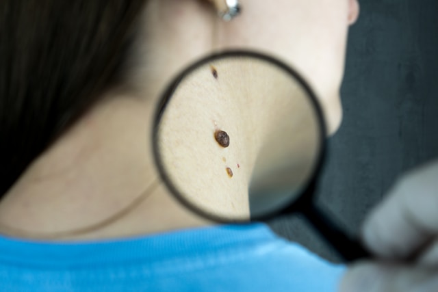
There are millions of skin cancer cases diagnosed around the world each year which makes early detection critical for successful treatment. But there are many advancements in technology that has transformed detection of skin cancer so that healthcareprofessionals are able to identify suspicious moles and lesions accurately.
You can ask your dermatologist about how often to check for skin cancer. This can differ according to your risk factors. A revolutionary aspect of dermatology is pattern recognition AI. This can greatly improve the accuracy of skin cancer detection. Dermascopic imaging and total body photographs will be used so that AI algorithms are able to analyse the images and identify if there is a change in your lesions or moles. If there is a suspiciouslesson, this will be benchmarked against a global dermascopic database. Healthcare professionals will be able to use AI so that they can understand the percentage of similar lesions that are malignant. This is great for early detection of skin cancer. This practice will highlight lesions that exhibit major changes over time. AI-powered laser-induced plasma spectroscopy or LIPS is used to analyse the biochemical composition of a suspicious skin lesion. This is a non-invasive technology. A LIPS score will be provided for the lesion and this will range between 0 and 1. When the score is higher, there is a higher risk of malignancy. This way, the healthcare professional will be able to evaluate the lesion further or carry out a biopsy. AI analysis can be used to improve diagnostic accuracy and lead to quick decision making.
RCM or reflectance confocal microscopy is an advanced imaging technique
Where you can have real time visualisation of skin structures at cellular resolution. This is a non-invasive technology and dermatologists are able to examine suspicious lesions at a microscopic level without having to carry out tissue biopsy. The skin layers can be reviewed in high resolution images and the dermatologist will be able to identify cellular features that are associated with skin cancer. This will improve the accuracy of the diagnosis and reduce the need to carry out invasive procedures. HFUS or high-frequency ultrasound can provide detailed visualisation of skin lesions and underlying structures. High frequency sound waves are used here so that they can penetrate the skin and produce high resolution images of tissue layers. The depth of skin lesions and theircharacteristics can be assessed this way. You can use this method to assess lesions in challenging locations on the body.
OCT or optical coherence tomography is another non-invasive imaging technique.
It will provide real time cross sections images of skin tissues. This way, the dermatologists will be able to check high resolution images of tissue microstructure so that cellular and subcellular features can be visualised clearly. This is great for evaluating lesions that have unclear clinical features. It is also good for monitoring the response of the patient to treatment. Another non-invasive imaging technique used to capture multiple images of the skin is multispectral imaging and skin lesions will be assessed on their spectral characteristics.


